0
CART
0 item
Categories
-
Veterinary Books
-
Companion Animals
- Anaesthesia/Analgesia
- Anatomy
- Cardiology
- Cytology/Laboratorial
- Dermatology
- Diagnostic imaging
- Emergency
- Endocrinology
- Endocrinology/ Reproduction
- Equine
- Ethology
- Infectious diseases and immunology
- Nephrology/Urology
- Nutrition
- Oncology
- Ophthalmology
- Parasites
- Pet owner educational atlas
- Rehabilitation
- Senior care
- Surgery
- Surgery and traumatology/Orthopaedics
- Veterinary Dentistry
- Livestock
- Management
-
Companion Animals
- Dentistry Books
- Medicine Books
- CE Webinar
- CE Webinar
- French books
- E-books


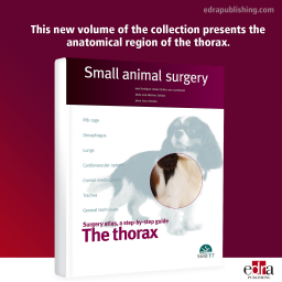

The Thorax. Small...
Price
CAD 154.35
This volume of the collection presents the anatomical region of the thorax. It gives a detailed description of thoracic surgical procedures, ranging from the most common ones, such as thoracotomy, to the most complex ones such as the placement of a transvalvular patch in cases of pulmonary valve stenosis.
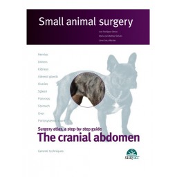

The cranial abdomen....
Price
CAD 162.75
Servet presents the third volume of the publication “Small animal surgery”, which describes the main surgical procedures on the cranial abdomen of the dog, cat and ferret. This work, which has been acclaimed by professionals, stands out for the excellence of its images and the step-by-step description of each surgical technique using real clinical cases.
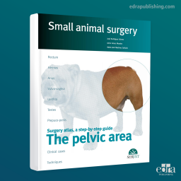
The Pelvic Area. Small...
Price
CAD 145.95
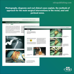
Photographs, diagrams and real clinical cases explain, step by step, the methods of approach and resolution for the main surgical interventions in the rectal, anal and perineal zones. An essential work for both practicing professionals and surgery students.
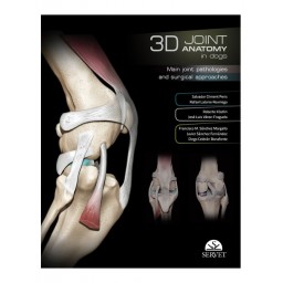
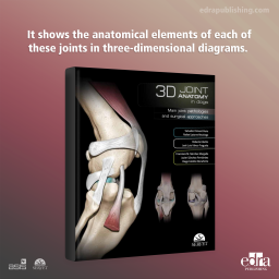
3D Joint Anatomy in...
Price
CAD 132.30
A visual guide with a strongly educational approach covering the main joints in the limbs of the dog. It shows the anatomical elements of each of these joints in three-dimensional diagrams. The views chosen for each case have been selected for a practical purpose, showing the position of the elements involved in the most commonly used surgical approaches. It also describes the key orthopaedic conditions affecting each joint and the most commonly used surgical approaches. It contains a large number of images and illustrations, and a selection of views presented in digital video format.