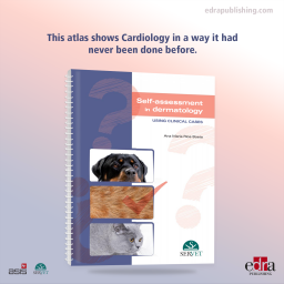
This atlas shows Cardiology in a way it had never been done before. The images it contains, of both the healthy heart and the main heart diseases that affect dogs and cats, have a unique technical and artistic value.
This atlas shows Cardiology in a way it had never been done before. The images it contains, of both the healthy heart and the main heart diseases that affect dogs and cats, have a unique technical and artistic value.
Author
Juan Carlos Jiménez de la Puerta
He graduated in Veterinary Medicine from the Complutense University of Madrid in 1978. He obtained his PhD in Veterinary Medicine in 1993. He obtained a diploma in vascular and nerve microsurgery from the University of Extremadura in 1987, as well as a diploma in microsurgery from the Gregorio Marañón Hospital in 1988. He received a scholarship from the Madrid College of Veterinary Surgeons to complete an internship at the Department of Medicine and Experimental Surgery of the Gregorio Marañón Hospital in 1988, and another from the Centre for Industrial Technological Development to work at the Department of Cardiovascular Pharmacology of ALTER Laboratories (1989-1992).
He obtained a diploma in Echocardiography from the University of Murcia in 1996. In 1998 he won the AVEPA prize for scientific photography and was awarded the Miguel Luera prize in 2000. He is the author of several publications and papers in national and international journals and congresses. He has worked as a clinical veterinary surgeon since 1981 and has specialised in cardiology since 1988. He has organized several scientific and artistic photography exhibitions.
Table of Contents
1. Heart anatomy
Orientation and location in the thoracic cavity
Size of the heart
Pericardium
Cardiac surface and main blood vessels
Fibrous skeleton
Right atrium
Right ventricle
Left atrium
Left ventricle
2. Mitral valve
Mitral valve annulus
Leaflets
Chordae tendineae
Papillary muscles
3. Tricuspid valve
Tricuspid valve annulus
Leaflets
Chordae tendineae
Papillary muscles
4. Aortic valve
Outflow tract of the left ventricle
Aortic root
5. Pulmonary valve
Annulus
Cusps
Commissures
Sinotubular junction
Subcommissural triangles
Sinuses of valsalva
6. Atrioventricular valve dysplasia
Mitral valve dysplasia
Tricuspid valve dysplasia
7. Aortic valve stenosis
8. Pulmonary valve stenosis
9. Patent ductus arteriosus
10. Septal communications
11. Myxomatous valve degeneration
Histopathology
Pathological changes
12. Dilated cardiomyopathy
13. Hypertophic cardiomyopathy
14. Pericardium and neoplasia
Pericardial effusion
Cardiac tumours
15. References
Data sheet
Specific References
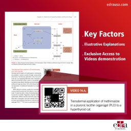
This book is unique, developed by the main worldwide experts in this issue. It is an extremely valuable tool for any veterinary practitioner willing to deepen into physiopathology, clinical signs, diagnosis, treatment and prognosis of feline endocrine diseases, with an approach and contents never published so far. the most common endocrine conditions in cats are deeply discussed and addressed: hyperthyroidism, type-II diabetes mellitus, and growth hormone excess.
Correct vaccination of dogs and cats requires consideration of a broad range of clinical situations and vaccination options, and obliges veterinary surgeons to constantly update their knowledge in order to appropriately deal with the challenges that arise in daily clinical practice. Using a thoroughly practical approach, this book takes an in-depth look at vaccines and vaccination to provide veterinary professionals with the information they require to address the many doubts and questions that arise in relation to this topic.
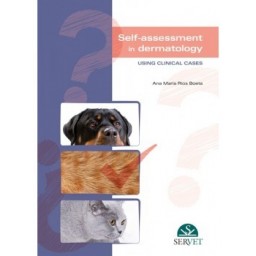
This self-assessment manual allows veterinary surgeons and veterinary students to test their knowledge of small animal dermatology. The book presents a variety of different clinical cases, replete with images of affected patients and relevant data. Based on this information, the reader can identify the disease process in question in a simple and entertaining way, thus refreshing and expanding their knowledge of diagnosis and treatment.
Cette deuxième édition, en plus d’offrir les bases de l’interprétation radiologique et du diagnostic des pathologies, élargit considérablement son contenu avec un chapitre sur les principes de la radiologie et un atlas de positionnement et d’anatomie radiographique, comportant des indications très claires et précises pour obtenir les meilleurs résultats lors du diagnostic radiologique.
This handbook about small animal emergency care contains the diagnostic and therapeutic approaches to the most common emergencies seen in small animal practice, classified by organ systems. For each emergency, a definition of the condition that has caused the situation, its causes, the diagnostic approach, and the most appropriate treatment is provided. The goal is to provide an easily accessible quick reference guide to save valuable time during stressful emergency medical cases.
This atlas shows Cardiology in a way it had never been done before. The images it contains, of both the healthy heart and the main heart diseases that affect dogs and cats, have a unique technical and artistic value.