
Mastitis represents a major endemic disease of dairy cattle, being a very complex disease that depends on several factors and usually presented subclinically in herds. The treatment and control entails high costs and losses for the industry. The author has developed an accurate, visual review analysing the most highlighted features of this condition and pointing out the main control measures to prevent it.
Mastitis represents a major endemic disease of dairy cattle, being a very complex disease that depends on several factors and usually presented subclinically in herds. The treatment and control entails high costs and losses for the industry. The author has developed an accurate, visual review analysing the most highlighted features of this condition and pointing out the main control measures to prevent it.
Authors:
MARIA E. PRADO
MV, PhD, DACVIM.
Dr. Maria E. Prado obtained her veterinary medicine degree (MV) from the University of Zulia in Maracaibo, Venezuela in 1987. Following two years of bovine private practice, she came to the United States to further her education. She completed a one year internship in Food Animal Medicine and Surgery and Equine Theriogenology at Oklahoma State University (1993-1994) followed by a residency in Food Animal Medicine and Surgery (1995-1998) and board certification in the American College of Veterinary Internal Medicine (2000). Dr. Prado performed graduate studies in Veterinary Pathobiology and obtained a PhD at Oklahoma State University in 2003 and a postdoc in Molecular Virology. Since 2005, she has been employed by the University of Tennessee where she conducts research in the areas of bovine mastitis and respiratory disease as well as teaching.
Key Features:
➜ Online visualisation of the sample chapter.
➜ Presentation brochure in PDF format.
➜ Author ́s CV.
➜ Sample chapter compatible with iPad.
Table of Contents:
1. The bovine mammary gland
Macroscopic anatomy
Microscopic anatomy
Physiology of the lactation
Mammary gland growth (mammogenesis)
Milk production and lactation
Milk ejection reflex
Defense mechanisms
Physical barriers
Cellular defenses
Noncellular defenses
2. Epidemiology
Prevalence and incidence
Distribution and frequency of isolation of
pathogens from clinical mastitis
Source and transmission of infection
Risk factors
3. Pathogenesis
Infectious aetiology
4. Clinical presentation
5. Diagnosis
Subclinical mastitis
SCC
Clinical mastitis
Physical exam
Milk culture and sensitivity
6. Treatment
7. Control and prevention
8. Case investigation
Herd goals
Mastitis problem investigation
9. References
Data sheet
Specific References
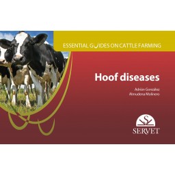
A visual and instructive book written by experts in the field, which shows readers how to analyse the most important characteristics of lameness and the main measures for its control and optimal prevention.
Atlas entirely dedicated to infectious bursal disease based on a practical and graphic approach of the topic. This handbook has been carried out by highly experienced authors closely involved with this disease in Africa, where its prevalence and incidence are significant. This updated work has been carefully developed including the most essential contents, so that it depicts a valuable tool to cope and control this virus as far as possible. Includes many helpful tips and visual graphic resources provided by the authors to enrich and turn this atlas into a reference in its field. Furthermore, diagnosis and vaccination have been thoroughly explained, highlighting vaccination programmes and types of vaccines. The atlas format helps to make the contents understandable for readers.
Manuel Fernández Sánchez, author of the best seller The Oestrus Cycle of the Cow. A Photographic Atlas, now brings us his second book Reproduction and ultrasound examination in cattle, published by Servet. It offers a detailed comparison of the uterine and ovarian structures in all phases of the oestrous cycle. This publication is characterized by an original introduction to both photographic and ultrasound images of the different anatomical structures and their pathological conditions. In this way, the author succeeds in simplifying the interpretation of reproductive diagnosis using ultrasound, by guiding the reader and enabling him to visualise the structures on which ultrasonography is performed.
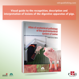
Visual guide to the recognition, description and interpretation of lesions of the digestive apparatus of pigs. Integrates macroscopic and microscopic findings associated with lesions that develop during the course of various pathological processes. Brief descriptions of the main anatomopathological features of each image are provided, which is essential for proper understanding of the disease process, diagnosis and underlying causes.
This book offers essential information about how to obtain samples for routine clinical and pathological investigations veterinarians habitually follow in pig farms. In addition, a complete and easy-to-perform necropsy procedure is shown. Picture galleries illustrate these procedures in a very easy way to follow. The guide also contains a set of pictures representing the main pathologies veterinarians can find in their practice. The guide is very visual and literature is reduced in order to facilitate the use of the book. This design, together with the book format and binding makes this guide very easy to use in practice by veterinarians and farm workers.
Mastitis represents a major endemic disease of dairy cattle, being a very complex disease that depends on several factors and usually presented subclinically in herds. The treatment and control entails high costs and losses for the industry. The author has developed an accurate, visual review analysing the most highlighted features of this condition and pointing out the main control measures to prevent it.